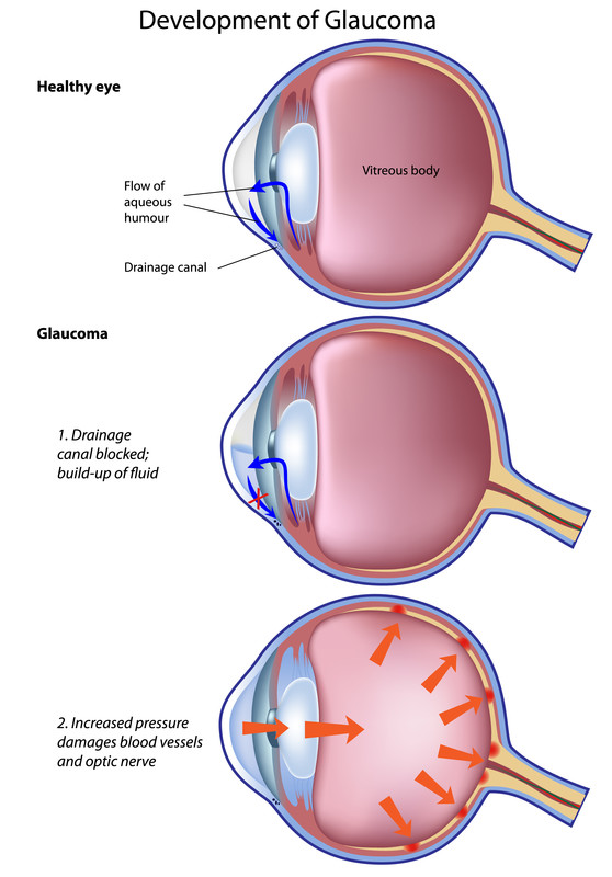
Glaucoma
Glaucoma is a group of diseases that damage the eye’s optic nerve and can result in vision loss and blindness. There is no cure for glaucoma. Vision lost from the disease cannot be restored.
Several large studies have shown that eye pressure is a major risk factor for optic nerve damage. When the optic nerve is damaged from increased pressure, open-angle glaucoma and vision loss may result. Another risk factor for optic nerve damage relates to blood pressure. Thus, it is important to also make sure that blood pressure is at a proper level by working with the medical doctor.
Whether one develops glaucoma depends on the level of pressure one’s optic nerve can tolerate without being damaged. This level is different for each person. Glaucoma can also develop without increased eye pressure.
Some people, listed below, are at higher risk than others:
- African Americans over age 40
- Everyone over age 60, especially Mexican Americans
- People with a family history of glaucoma
In some people with certain combinations of these high-risk factors, medicines in the form of eyedrops reduce the risk of developing glaucoma by about half.
At first, open-angle glaucoma has no symptoms. It causes no pain. Vision stays normal. Glaucoma can develop in one or both eyes.
Without treatment, people with glaucoma will slowly lose their peripheral (side) vision. As glaucoma remains untreated, people may miss objects to the side and out of the corner of their eye. They seem to be looking through a tunnel. Over time, straight-ahead (central) vision may decrease until no vision remains.
Glaucoma is detected through a comprehensive dilated eye exam that includes the following:
- Visual acuity test. This eye chart test measures how well the person sees at various distances.
- Visual field test. This test measures the person’s peripheral (side vision). It helps the eye care professional tell if the person has lost peripheral vision, a sign of glaucoma.
- Dilated eye exam. In this exam, drops are placed in the eyes to widen, or dilate, the pupils. The eye care professional uses a special magnifying lens to examine the retina and optic nerve for signs of damage and other eye problems. After the exam, the close-up vision may remain blurred for several hours.
- Tonometry is the measurement of pressure inside the eye by using an instrument called a tonometer. Numbing drops may be applied to the eye for this test. A tonometer measures pressure inside the eye to detect glaucoma.
- Pachymetry is the measurement of the thickness of the cornea. The eye care professional applies a numbing drop to the eye and uses an ultrasonic wave instrument to measure the thickness of the cornea.
Vision screening is an efficient and cost-effective method to identify adults with visual impairment or eye conditions so that a referral can be made to an appropriate eye care professional for further evaluation and treatment.
Source: The National Eye Institute
SUGGESTED VISION SCREENERS INCLUDE:
- Includes contrast sensitivity tests (sine-wave gratings)
- 4 lighting conditions – Day and night, with and without glare
- Meets ANSI standards with unsurpassed homogeneous illumination
